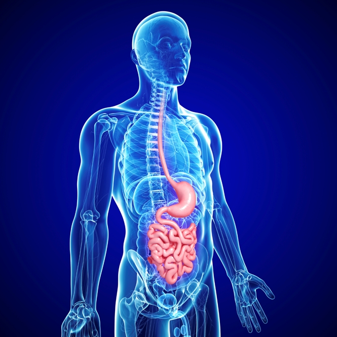
New Light on the Treatment of Spinal Tumors
Malignant tumors of the spine account for up to 8% of all tumors found in the central nervous system. The diagnosis of spinal tumors is routinely established according to WHO criteria, and the most common tumors are meningiomas, nerve sheath tumors, ependymomas, and astrocytomas. These tumors typically grow within the spinal canal but outside of the nerves. Fortunately, they tend to be slow-growing and benign, though they can cause some pain and weakness.
Currently, the conventional treatment of choice for most spinal tumors is total surgical removal, with neurosurgeons doing their best to preserve the patient’s neurological function. However, it is not uncommon for neurosurgeons to be unable to remove all of the spinal tumor mass. In some cases, the surgeon can clearly see that some of the tumor still remains. In other cases, however, some microscopic spots (or foci) of cancer remain that are not detectable or clearly seen by the surgeon. This is known as subtotal resection (STR). With STRs, which are fairly common with astrocytomas, tumor recurrence is a frequent consequence. Such recurrences are thought to take place in about 70% of STR cases postoperatively.
A common reason for the incomplete surgery is that the surgery must be ended prematurely due to the patient’s declining neurological functioning during the operation. Another key reason is that the remaining tumor tissue in the brain cavity is generally not visible under the conventional microscope. For these reasons, a more clinically reliable technique for visualizing the spinal tumor tissue is needed during surgery.
This is where fluorescence-guided surgery can enter the picture. The photosensitizer known as 5-aminolevulinic acid (5-ALA) leads to accumulations of the natural compound called protoporphyrin IX, with the substance accumulating preferentially in brain tumors and spinal tumors. Upon exposure to light, the tumor tissue will fluoresce or glow, thus clearly revealing itself to the neurosurgeon.
The use of 5-ALA as part of fluorescence-guided surgery for malignant brain tumors has led to a higher rate of complete surgery of malignant glioma when compared to conventional “white light” procedures. Furthermore, 5-ALA has been used for the visualization of other brain tumors such as meningiomas and metastases as well. Recently, the use of 5-ALA was also reported in spinal tumors, with fluorescence being observed in a few cases of ependymomas, meningiomas, and malignant gliomas.
Researchers at the Medical University of Vienna (Austria) recently sought to determine which spinal tumor types could be visualized with the 5-ALA–induced fluorescence method in a series of 52 patients with various spinal cancers. Surgery was performed in 50 of 55 cases; the remaining five cases underwent biopsy. Oral solutions of 5-ALA were administered three hours before beginning anesthesia in all patients. Upon exposure to violet-blue light, the fluorescence status in each tumor was checked, and the specific fluorescence pattern was analyzed.
What follows are the main findings from this study:
- Complete surgical removal was achieved in 37 of the 55 cases
- Incomplete or partial surgical removal took place in 13 cases; 5 of these cases involved STR.
- Protoporphyrin IX fluorescence was visible in 55% the cases.
- Fluorescence was mainly detected in 100% of ependymomas, meningiomas, hemangiopericytomas, and in drop metastases of primary CNS tumors.
- In contrast, none of the neurinomas, carcinoma metastases, and primary spinal gliomas showed fluorescence.
- Residual tumor foci or spots were detected by fluorescence and tne removed surgically in four out of eight ependymomas despite the neurosurgeons’ initial assumption that the tumor had been completely removed.
Thus, the 5-ALA-induced fluorescence technique helped identify various types of spinal tumors, and this proved to be a useful tool for the detection of potential residual tumor foci in ependymomas.
“This combination of treatment has the purpose of better overall survival for some cancer patients by positively influencing local control,” the authors wrote. “The role of surgical intervention in the context of managing cancer patients is continuously being redefined for this challenging population. The modern surgeon must stay abreast of all advances in their field in order to provide the best care for their patients.”
Support us by buying our book, The Medicine of Light, and ebooks from our Photoimmune Discoveries eBook Series.
Sources
Millesi M1, Kiesel B, Woehrer A, Hainfellner JA, Novak K, Martínez-Moreno M, Wolfsberger S, Knosp E, Widhalm G. Analysis of 5-aminolevulinic acid-induced fluorescence in 55 different spinal tumors. Neurosurg Focus. 2014 Feb;36(2):E11.
Araujo JL, Veiga JC, Figueiredo EG, Barboza VR, Daniel JW, Panagopoulos AT. Management of metastatic spinal column neoplasms – an update. Rev Col Bras Cir. 2013 Dec;40(6):508-514.
Wu L, Deng X, Yang C, Xu Y. Spinal intradural malignant peripheral nerve sheath tumor in a child with neurofibromatosis type 2: the first reported case and literature review. Turk Neurosurg. 2014;24(1):135-9.
© Copyright 2014, Photoimmune Discoveries, BV