During treatment with photodynamic therapy, the photosensitizer accumulates more strongly in diseased than in normal tissues, and this helps explain PDT’s ability to selectively target tumors. The accumulation of the photosensitizer can also result in fluorescence, or a colorful glow that is emitted by the diseased or malignant tissue. The difference in photosensitizer concentrations in normal versus diseased tissues can produce contrasting colors between the tissues, enabling the physician to see evidence of the disease beyond a clearly visible tumor.
This diagnostic approach is known as Photodynamic Diagnosis, or PDD (also called photodiagnosis or fluorescence diagnosis). German scientists first recognized the value of PDD for detecting abnormal growths back in the early 1900s. The “glowing” of the tissue is due to the effect of a photosensitizer interacting with light. Chemists and physicists like to describe the light-triggered activation of the photosensitizer as “excitation by a specific wavelength of light within the visible spectrum range”.
A classic example of how PDD can be used to help detect cancer involves basal cell carcinoma, the most common form of cancer worldwide. Upon exposure to ultraviolet light, the cancerous areas shows a coral red fluorescence that can clearly be distinguished from the much lower fluorescence of adjacent normal tissue. Other forms of non-melanoma skin cancer can also be diagnosed in this way.
Using PDD to Catch Cancer Early
Many cancers are first detected only after they’ve become bothersome or symptomatic. Unfortunately, by that time, they’ve had ample opportunity to evolve into a more established or aggressive disease. Had these malignancies been detected much sooner, they could have been treated far more effectively, resulting in better long-term survival.
PDD can detect very tiny tumors as well as clusters of cancer cells as well as abnormal cells (dysplastic tissue). It is thus particularly helpful in identifying and localizing precancerous conditions and early cancerous lesions—in other words, catching cancer in its earliest phases of evolution, before it has a chance to become a life-threatening condition.
Let us consider the example of skin cancer. Actinic keratosis is a common skin condition caused by excessive sun exposure. This condition can precede the development of squamous cell carcinoma. A study out of Radboud University Nijmegen Medical Centre in Nijmegen, the Netherlands, recently found that PDD could help reveal various stages of malignant development from actinic keratosis to squamous cell carcinoma.
In essence, the Dutch researchers were able to show that variations in the intensity of fluorescence (the brightness of the “glow”) seemed to reflect the degree of invasiveness and cell abnormalities that could help predict the development of a more aggressive skin cancer later on. It follows that PDD may serve as an important diagnostic and monitoring tool in clinical practice.
Making Surgery and PDT More Effective for the Long Term
PDD is also very helpful in cases where the surgeon cannot see the tissue that needs to be excised. This is known as the surgical margin, and it’s a major reason that the success of surgery can be short-lived. By using PDD, the surgical margin can be more accurately seen. This enables what is now known as fluorescence-guided surgery—an especially attractive approach for brain tumors and certain other cancers that pose immense challenges for conventional cancer surgery.
At the same time, it should be emphasized that photodynamic therapy, or PDT, is often a great option for these early-stage situations, and it works very logically as a complement to PDD. What you see —thanks to the fluorescence effect during PDD—is literally what you are able to treat directly using PDT.
In summary, photodiagnosis is a great clinical tool that has two very broad objectives:
- detecting cancerous, precancerous and other diseased tissues
- supporting image-guided therapy (surgery and/or PDT)
More specifically, in clinical practice, the use of PDD is focused on these five targets:
- detection of precancerous changes in order to assist with cancer prevention
- detection of cancerous tissue in the early stages, thus enabling fast, early treatment
- detection of recurrence of the cancer, again at any early stage
- for monitoring the disease, to see if the cancer returns, spreads or progresses
- helping to possibly exclude the presence of cancerous disease.
Advantages and Applications of PDD
A report in the July 2013 issue of the medical journal, OncoTargets & Therapy, describes PDD as a “fast, easy, noninvasive, selective, and sensitive diagnostic tool for estimation of treatment results in oncology.” The article goes on to describe this diagnostic method as “very sensitive and specific especially in diagnostics of small changes that are not visible using white light endoscopic procedures.” In other words, PDD enables physicians to actually see what it is they’re supposed to be treating!
Let’s consider the example of gastrointestinal tumors or adenocarcinomas of the digestive tract. You’re more at risk of developing such tumors if you have a history of Barrett’s esophagus, adenomatous polyps, or long-standing ulcerative colitis. If you’re judged to be in such a risk category, you may undergo annual or biannual endoscopies with multiple biopsies—that would be the conventional strategy
The problem with these conventional methods is that most of the areas of cell abnormality (dysplasia, precancer) are either very obscure or impossible to see with standard endoscopy methods. Even the practice of tissue staining (chromoendoscopy) can yield very mixed results. Again, PDD could offer a meaningful and effective solution to this problem.
Many applications of PDD have been identified. What follows is a list of some of the applications in the realm of oncology:
- Basal Cell Carcinoma. PDD appears to be a useful tool for detecting superficial basal cell carcinoma as well as for predicting treatment response. In some studies, the degree of fluorescence of the lesions prior to PDT has been shown to predict a complete treatment response. Nevertheless, the study findings have been inconsistent, and research is needed to better assess PDD’s usefulness, ideally using Bremachlorin-PDD followed immediately by Bremachlorin-PDT (the “see and treat” photodynamic strategy).
- Bladder Cancer. Many well-designed studies have shown that PDD significantly improves the detection of bladder cancer when compared with standard white-light cystoscopy. PDD also enables more complete surgical treatment, resulting in a lower residual disease and thus lower recurrence rates, as reported in the November 2013 issue of European Urology. Studies are now needed using Bremachlorin-PDD followed immediately by a combination of surgery and PDT. PDD can then be used as a follow-up monitoring tool to check for early signs of a possible recurrence.
- Brain Cancer. PDD has been successfully used to help guide the surgical treatment of brain tumors, enabling the surgeon to minimize damage to the normal, healthy brain tissues. The combination of PDD with fluorescence-guided surgery and intraoperative PDT (“to see and to treat”) offers a powerful approach to the treatment of malignant brain tumors, as reported in a 2010 issue of Methods in Molecular Biology..
- Cervical Cancer (and other female cancers. Scientists are currently trying to assess whether PDD could be useful in gynecologic conditions such as cervical intraepithelial neoplasia (CIN), vulvar intraepithelial neoplasia (VIN), endometriosis, breast cancer and ovarian cancer. With regard to cervical cancer, the use of a PDD-based “cervical smear” may hold promise for detecting early signs of cancer, as reported in the May 2012 issue of Photomedicine and Laser Surgery.
- Esophageal Cancer. Barrett’s esophagus is a condition that can increase the risk of esophageal cancer, a very deadly disease. The non-dysplastic form of Barrett’s esophagus is not likely to result in cancer, while there is a significant risk of cancer with the dysplastic form. Unfortunately, it is difficult for the pathologist to differentiate non-dysplastic Barrett’s esophagus from Barrett’s esophagus with low-grade dysplasia. This is where PDD seems to have strong efficacy, as reported in the July 2012 issue of Lasers in Surgery and Medicine.
Other studies have suggested the potential usefulness of PDD for early detection of breast cancer, lung cancer, ovarian cancer, and colon cancer, and head and neck cancers.

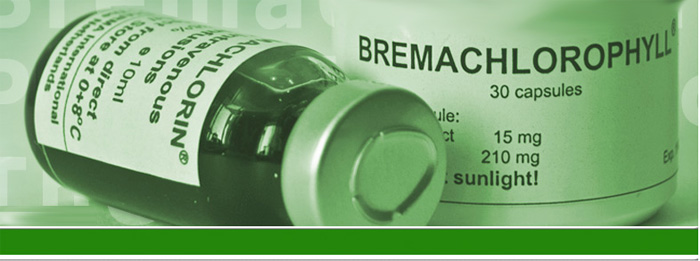
Optimizing PDD with Bremachlorin®
In order for photo diagnostic methods to work, they must incorporate the photosensitizing agents. We now know that the best photosensitizers for tumor detection show a high fluorescence yield, high selectivity for cancerous tissue during the first 24 hours after administration, and rapid elimination from the body (to minimize photosensitivity). Many so-called second-generation photosensitizers are beginning to show promise in this regard. These agents have a low risk of side effects, are easy to administer, and seem to be diagnostically useful because they accumulate so easily in tumor tissues.
Bremachlorin® meets all of the requirements for the best photodiagnostic agent and bas been proven clinically to aid in the detection of many types of malignant tumors. Simply by illuminating the diseased tissue Bremachlorin® serves a very helpful role. At the same time, with the right light intensity, Bremachlorin® can be used as part of a very effective PDT regimen to treat the cancer on the spot.
By detecting precancerous and cancerous growths, Bremachlorin-PDD provides vital information for anyone hoping to avoid a malignancy. This information can be used to help alert you, the patient, to the need to be more aggressively proactive in your efforts to ward off cancer, perhaps using a combination of nutritional, herbal and innovative medical strategies. As a novel method of tumor detection, Bremachlorin-PDD could very well play a central role in the medicine of the future—an approach grounded in the authentic, biologically guided practice of preventive medicine.
Certainly in the context of surgery, PDD has invaluable power. When a surgeon is preparing to remove some cancerous tissue, it is very helpful to see the margins or border of malignant tissue Thus, PDD proc an be used for planning the surgical procedures. It can also be used after photodynamic therapy at a follow-up visits for the visualization of the therapeutic outcome.
Support us by buying our book, The Medicine of Light, and ebooks from our Photoimmune Discoveries eBook Series.


 English
 Français
 Deutsch
 Nederlands